

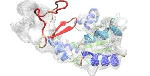
Proteins are one of the major building blocks of life, and they carry out many chemical reactions related to life. This includes how the body interacts with disease-causing agents. Understanding and treating diseases requires scientists to determine the structures and shapes of proteins involved in these interactions.
Three-dimensional computer modeling and electron microscopy are two of the tools scientists are currently using to help learn more about these proteins. To spur development of more effective tools, EMDataResource, a global organization for the storage and retrieval of three-dimensional electron microscopy data, sponsored the 2019 Cry0-EM Map-based Model Metrics Challenge. The goals of this challenge were to assess the quality of models that can be produced using current modeling software; check the reproducibility of modeling results from different software developers and users; and compare the performance of current metrics used for evaluation of models.
Abhishek Singharoy, researcher with the Arizona State University Biodesign Center for Applied Structural Discovery and assistant professor with ASU’s School of Molecular Sciences, led the CryoFold team. Singharoy’s group was recognized in the challenge for doing very well in both lower-resolution and higher-resolution targets. CryoFold is an Ab-initio modeling platform interfacing between three popular software tools such as MAINMAST, developed by Dr. Genki Terashi and Professor Daisuke Kihara at Purdue University, Modeling Employing Limited Data (MELD) developed by Dr. Alberto Perez, now at University of Florida and Professor Ken Dill, Stony Brook University, and Resolution Exchange Molecular Dynamics Flexible Fitting (ReMDFF) developed by Dr. Singharoy and Professor Klaus Schulten, University of Illinois at Urbana-Champaign.
Singharoy noted: “The work assessed by members of Protein Data Bank (PDB) and Electron-Microscopy Data Bank (EMDB) highlights the importance of integrative modeling in the fields of structural biology, biophysics and biochemistry. It is very encouraging for our team members to see the excellent performance of molecular dynamics flexible fitting (MDFF) methods in this competition.”
“It is exciting to see the advances and successes coming from combining cryoEM maps and computational tools to provide atomistic detail of proteins. Solving the protein structure is crucial to understanding how they work and to design new drugs that target them,” said Perez.
A paper on the results of the competition is published in the journal Nature Methods.
Singharoy offered, “Understanding three-dimensional protein structures will help us tackle many chronic and fatal diseases associated with proteins.”