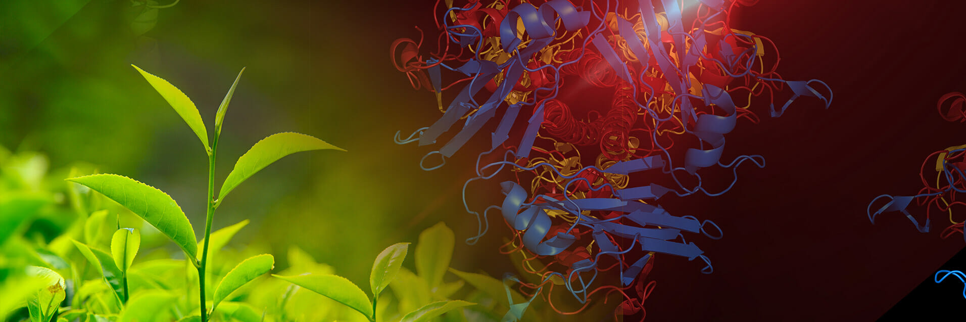
Cryogenic electron microscopy (Cryo-EM) is a powerful technique capable of penetrating the mysteries of the molecular world at near atomic resolution. Along with X-ray crystallography and nuclear magnetic resonance spectroscopy (NMR), cryo-EM has been a boon to the field of structural biology, helping to unlock the fine details of proteins and their binding activities with small molecules. The enormous power and versatility of the method earned cryo-EM researchers the 2017 Nobel Price in Chemistry.
Instead of light, cryo-EM uses a beam of electrons for imaging. The technique holds enormous potential for drug discovery, as cryo-EM can reveal the subtle binding activities of drug molecules with their target proteins, particularly larger biomolecules that are resistant to crystallization. Cryo-EM relies on embedding molecules in vitrified water that has been chilled with ethane, creating a glasslike cocoon around the sample.
An electron beam is used to produce thousands of images of sample biomolecules, which are then fitted together using complex algorithms and eventually translated into a crisp, high resolution 3D structure. One significant hurdle for cryo-EM lies in handling the enormous volume of data generated from multiple, fuzzy sample images in different orientations and assembling these into a final structure. Limitations in data processing have made detailed investigations of protein-drug binding interactions challenging and time consuming.
In the new study, researchers borrow techniques that have been successfully applied to X-ray crystallography, specifically flexible fitting models and quantum mechanics/molecular mechanics (QM/MM) methods. According to lead author John Vant, “this is the first attempt at combining quantum mechanics calculations with a flexible fitting protocol for cryo-EM.” Vant is a researcher in the Biodesign Center for Applied Structural Discovery and ASU's School of Molecular Sciences.
Co-author Rowley describes how further refinement of cryo-EM raw data is accomplished using neural network potential models (NNP) to model the bound drug molecule. “These models are simple to deploy in an automated workflow, so in combination with molecular dynamics flexible fitting, they give a general route to high-resolution protein-ligand complexes from low-resolution cryo-EM data.”
The innovations could help cryo-EM peer deeply into membrane proteins—critical drug targets that remain resistant to imaging via X-ray crystallography. In addition to static structures, the new techniques hold the promise of capturing crucial molecular dynamics.
The work recently appeared in the Journal of Chemical Information and Modelling.
Lead author John Vant was recently awarded a National Science Foundation Graduate Research Fellowship and the LeRoy Eyring Memorial Fellowship. He is joined by his colleagues from the Biodesign Center for Applied Structural Discovery and ASU’s School of molecular Sciences (Abhishek Singharoy, Daipayan Sarkar, Sumit Mittal and Barbara H. Munk) as well as Shae-Lynn J. Lahey and Christopher Rowley; Department of Chemistry, Memorial University of Newfoundland; Kalyanashis Jana and Ulrich Kleinekathofer, Department of Physics and Earth Sciences, Jacobs University Bremen; Mrinal Shekhar, Center for Development of Therapeutics, Broad Institute of MIT. (Sumit Mittal is now with VIT Bhopal University.)